OCEAN'S SILENT CONDUCTORS: UNRAVELING THE SECRET OF MICROBIAL FILAMENTS

 |
| Microscopic image of a cable bacteria filament |
Based on biogeochemical observations, it was suggested that cells within an individual filament displays two types of metabolism: anodic cells showing sulfide oxidation in anoxic zone and cathodic cells showing oxygen reduction in oxic zone by the electrons produced by the sulfide oxidation which gets transported along the filament.
 |
| General overview of electron transport from suboxic to oxic region by cable bacteria |
16S rRNA analysis shows that the cable bacteria features the core genome of “Desulfobulbaceae” with only 25 out of 806 core gene families not detected and comparison with other members of families revealed that genes involved in energy production and conversion, amino acid and carbohydrate transport and metabolism were underrepresented which indicates limited organotrophic catabolic potential.
The current model of cable bacteria metabolism is that, anodic sulfide oxidation in anoxic cells coupled with long distance electron transport (LDET) to cathodic oxygen reduction in oxic cells. Cable bacteria lack genes coding for Sox pathway, reverse-type dissimilatory sulfite reductase (rDSR), or flavocytochrome C sulfide dehydrogenase present normally in sulfide-oxidizing bacteria and encodes genes of the canonical sulfate reduction (DSR) pathway. The cable bacteria reverse this pathway for sulfate oxidation. The first step is production of sulfur by type III sulfide:quinone oxidoreductase (SQR) in periplasm which chemically reacts with sulfide to form dissolved polysulfide. Polysulfide gets reduced to sulfide by membrane-anchored polysulfide reductase (PSR) using electrons from quinone pool in cytoplasmic membrane. In the second step, sulfur formed by SQR is oxidized to sulfate by DSR which gets transported across the membrane by homologs of membrane protein YeeE, a cytoplasmic rhodanese and sulfur transferase TusA. Reduced sulfur from TusA reacts with DsrC to form DsrC-trisulfide (DCT) which is oxidized by bisulfite reductase DsrAB to release sulfite and DsrC. Sulfite is then oxidized to sulfate by adenosine-5-phosphosulfate reductase (AprAB) and sulfate adenylyltransferase (Sat), generating ATP by substrate level phosphorylation. Then sulfate is transported out of the cell by SulP. The electrons released by the oxidation of reduced DsrC and sulfite are transferred to the quinone pool in the cytoplasmic membrane by the DsrMK and QmoABC membrane complexes, respectively.
 |
| Metabolic processes of cable bacteria |
 |
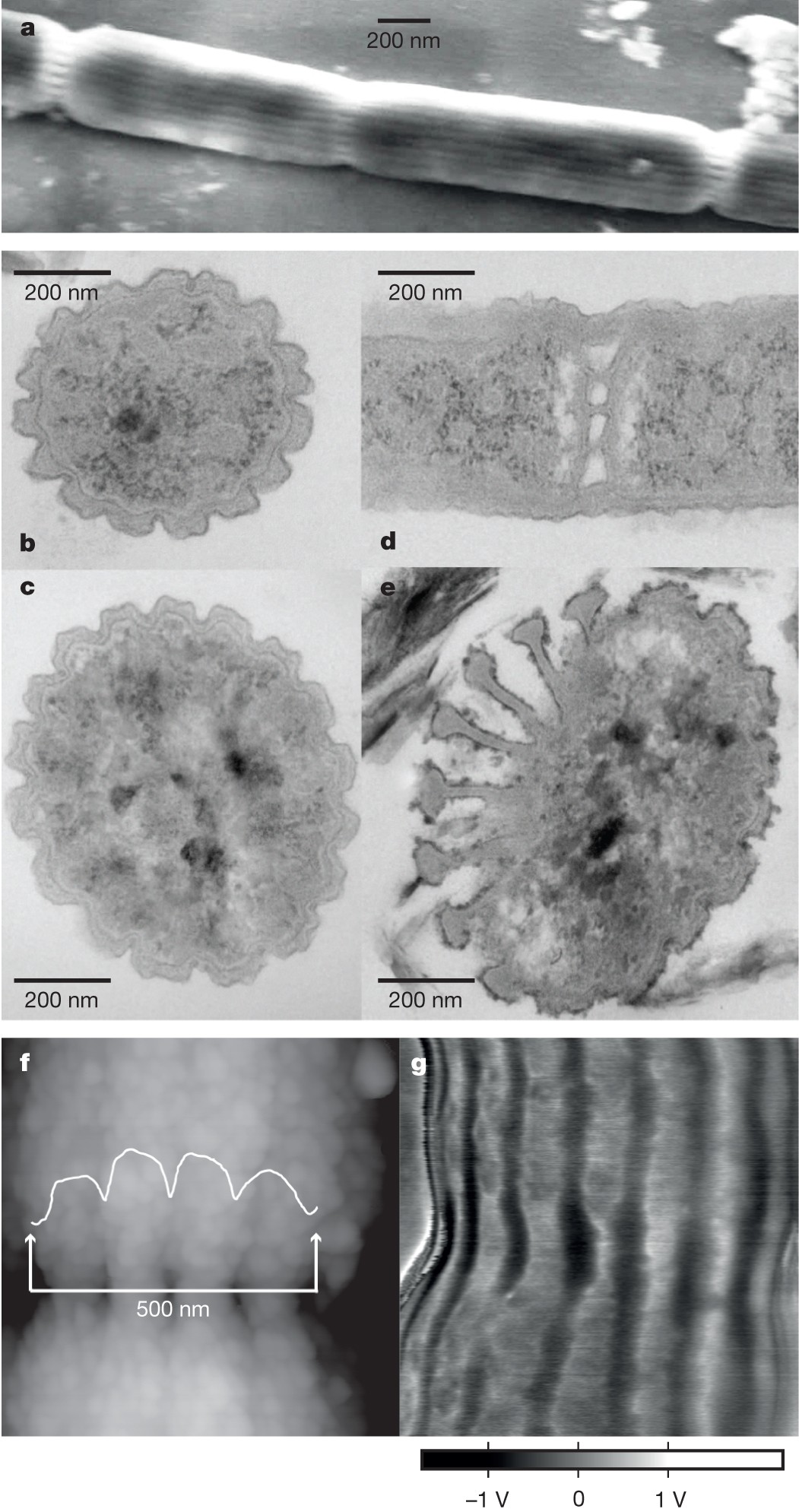
| AFM, SEM, TEM, and EFM micrographs of filamentous cable bacteria |
The filaments have a unique structure with uniform ridges along their entire length with 15 edges with each 400 nm wide or 17 edges with each 700 nm wide and adjacent cells within the filament separated by 200 nm covered by the outer membrane.
HAADF-STEM, AFM-IR, and ToF-SIMS data suggests that the conductive fibers are made of protein, present in a regular parallel pattern and rest upon a basal sheath rich in acidic polysaccharide most likely made of periplasmic peptidoglycan layer. Raman microscopy of intact cable bacteria confirmed the presence of cytochrome heme groups which was previously elucidated but also revealed the presence of cofactor with a ligated metal group. Raman spectroscopy of the conductive periplasmic fibers revealed that conduction of electrons does not involve cytochromes and suggested that the metal group is ligated with sulfur. The STEM-EDX and LEXRF data suggested that nickel is the metal coordinated with sulfur which was further supported by the ToF-SIMS.

These data provides the information on the model of the conductive fibers: fibers are made of protein embedded in a polysaccharide rich layer which holds the fibers together and adds tensile strength to it. The fibers are made of two domains: the central core contains nickel rich protein material surrounded by a thin Ni deficient protein layer resembling a normal electrical wire. The transfer of electrons through the insulating Ni deficient layer is mediated by periplasmic cytochromes.
This unique microorganism adds intricacy to the already diverse microbial world with each and every microorganism showing unique approaches to adapt to the environment. This also leads to a prospect of technological application with the discovery of the new conductive fibers providing a novel biomaterial with high conductivity for the application in bio-electronics. The discovery and study of such organisms provides a glance over nature's ingenuity and a path towards innovation that bridges biological and technological worlds for further breakthroughs in science and technology.
Kjeldsen, Kasper & Schreiber, Lars & Thorup, Casper & Boesen, Thomas & Bjerg, Jesper & Yang, Tingting & Dueholm, Morten & Larsen, Steffen & Risgaard-Petersen, Nils & Nierychlo, Marta & Schmid, Markus & Bøggild, Andreas & van de Vossenberg, Jack & Geelhoed, Jeanine & Meysman, Filip & Wagner, Michael & Nielsen, Per & Nielsen, Lars Peter & Schramm, Andreas. (2019). On the evolution and physiology of cable bacteria. Proceedings of the National Academy of Sciences. 116. 201903514. 10.1073/pnas.1903514116.
Pfeffer, Christian & Larsen, Steffen & Song, Jie & Dong, Mingdong & Besenbacher, Flemming & Meyer, Rikke & Kjeldsen, Kasper & Schreiber, Lars & Gorby, Yuri & El-Naggar, Mohamed & Leung, Kar Man & Schramm, Andreas & Risgaard-Petersen, Nils & Nielsen, Lars Peter. (2012). Filamentous bacteria transport electrons over centimetre distances. Nature. 491. 10.1038/nature11586.
Boschker, H. & Cook, Perran & Polerecky, Lubos & Thiruvallur Eachambadi, Raghavendran & Lozano, Helena & Hidalgo-Martinez, Silvia & Khalenkow, Dmitry & Spampinato, Valentina & Claes, Nathalie & Kundu, Paromita & Wang, Da & Bals, Sara & Sand, Karina & Cavezza, Francesca & Hauffman, Tom & Bjerg, Jesper & Skirtach, Andre & Kochan, Kamila & McKee, Merrilyn & Meysman, Filip. (2021). Efficient long-range conduction in cable bacteria through nickel protein wires. Nature Communications. 12. 3996. 10.1038/s41467-021-24312-4.
IMAGE SOURCES:
https://www.drugtargetreview.com/wp-content/uploads/shutterstock_1042543318-750x450.gif
https://asm.org/ASM/media/Article-Images/2022/July/Cable-bacteria_embed_2.jpg


Comments
Post a Comment