INTRATUMORAL MICROBIOTA: FROM HIDDEN PASSENGERS TO THERAPEUTIC ALLIES IN CANCER

Cancer has traditionally been viewed as a disease driven by genetic mutations, environmental factors, and dysregulation of cellular signalling pathways. However, recent advances in cancer research have revealed a previously overlooked component of the tumor microenvironment, the intratumoral microbiota. Once thought to be sterile, tumors are now known to harbor diverse communities of bacteria, fungi, and other microorganisms within their tissues. These microbial populations have been identified across a wide range of cancers, including colorectal, breast, lung, pancreatic, ovarian, and brain cancers, suggesting that their presence is not identical but may represent an important aspect of tumor biology.
The discovery of intratumoral microbiota has fundamentally changed our understanding of the tumor microenvironment. Rather than being passive inhabitants, these microorganisms actively interact with cancer cells, immune cells, and stromal components. Depending on their composition, intratumoral microbes can either promote tumor progression through inflammation, immunosuppression, DNA damage, epigenetic modifications, metabolic reprogramming, and chemoresistance, or contribute to antitumor immunity by stimulating immune responses, promoting anti-tumor signalling, secreting antitumour metabolites, and enhancing therapeutic efficacy. As a result, the intratumoral microbiota has emerged as a new frontier in cancer research, offering both challenges and opportunities for cancer treatment.
 |
| Tumor-promoting effects of intratumoral microbiota |
A central question arising from this discovery is how microorganisms are able to colonize tumors in the first place. Current evidence suggests several possible routes of microbial entry. In cancers associated with mucosal surfaces, disruption of protective barriers can allow microorganisms to escape their normal niches and infiltrate tumor tissues. For example, alterations in the intestinal mucosal barrier during colorectal cancer can increase permeability, facilitating bacterial translocation from the gut into surrounding tissues and even distant organs. Similarly, microorganisms can invade tumors from adjacent normal tissues that possess their own resident microbiota. Studies have demonstrated that the microbial composition of many tumors closely resembles that of neighboring healthy tissues, suggesting that local microbial communities may act as reservoirs for tumor colonization. Microorganisms may also reach tumors through hematogenous dissemination. Certain bacteria originating from the oral cavity or gastrointestinal tract can enter the bloodstream and preferentially accumulate in tumors at distant anatomical sites. Tumor-associated abnormalities in blood vessels, combined with local immune suppression, can facilitate the survival and persistence of these microbes within tumor tissue. In addition, emerging evidence suggests that some microorganisms may travel along neural pathways, particularly in the case of brain tumors, although the significance of this route remains under investigation. The ability of microorganisms to colonize tumors is not solely dependent on their route of entry. The tumor microenvironment itself provides conditions that favor microbial survival. As tumors grow, they often develop regions of hypoxia, necrosis, abnormal vasculature, and impaired immune surveillance. These conditions create ecological niches that can support microbial persistence and growth. In many cases, anaerobic and facultative anaerobic bacteria preferentially accumulate in the hypoxic and necrotic cores of tumors, where immune cell access is limited, and nutrient availability can support microbial metabolism.
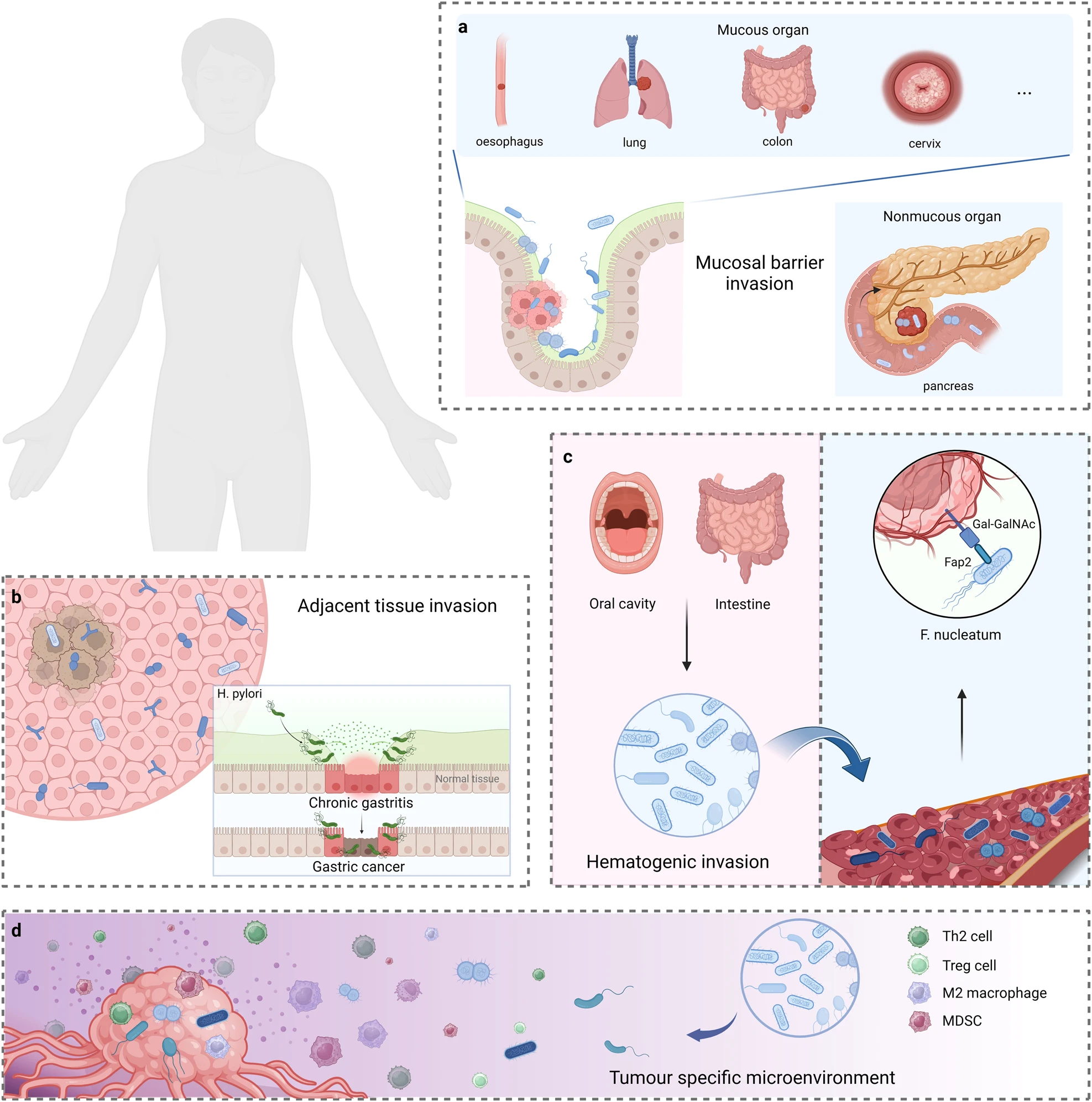 |
| Sources of Intratumoral Microbiota |
Interestingly, the same characteristics that enable microorganisms to colonize tumors also make them attractive candidates for cancer therapy. Unlike conventional drugs, which frequently struggle to penetrate the dense tumor microenvironment, certain bacteria can naturally seek out and accumulate within tumor tissue. This unique tumor-homing capability has inspired researchers to explore microorganisms as living therapeutic platforms capable of delivering anticancer agents directly to tumors while minimizing systemic toxicity.
One of the most promising applications of this concept involves the integration of tumor-colonizing bacteria with cancer immunotherapy. Chimeric Antigen Receptor T-cell (CAR-T) therapy has revolutionized the treatment of hematological malignancies but has achieved limited success against solid tumors. A major obstacle is the lack of suitable tumor-specific antigens and the inability of CAR-T cells to efficiently infiltrate and function within the immunosuppressive tumor microenvironment. Recent advances suggest that engineered tumor-colonizing bacteria may help overcome these limitations by selectively accumulating within tumors and locally delivering molecules that enhance CAR-T cell activity.
The researchers engineered the probiotic strain Escherichia coli Nissle 1917 (EcN), a bacterium widely recognized for its safety and previously explored in bacterial cancer therapy. The bacteria were equipped with a synchronized lysis circuit that allows them to selectively grow within the tumor microenvironment and periodically release therapeutic payloads. Rather than delivering conventional drugs, the engineered bacteria produced a synthetic protein tag consisting of superfolder green fluorescent protein (sfGFP) fused to a heparin-binding domain. This design enabled the released proteins to anchor to extracellular matrix components that are abundant in most solid tumors, effectively coating the tumor with an artificial target that could be recognized by GFP-specific CAR-T cells. By creating a target directly within the tumor microenvironment, the system bypassed the need for naturally occurring tumor antigens, one of the major obstacles limiting CAR-T therapy in solid cancers.
 |
| Overview of the ProCAR platform |
The researchers first investigated whether the synthetic tags could activate CAR-T cells. They observed robust activation when CAR-T cells encountered matrix-bound tags, whereas soluble GFP molecules generated only weak responses. The activated CAR-T cells exhibited increased expression of activation markers and produced higher levels of key inflammatory cytokines, including interferon-γ and TNF-α. Microscopy experiments further revealed clustering of CAR receptors at the interface between T cells and tumor cells, indicating successful immune synapse formation. Most importantly, the tagged tumors became susceptible to CAR-T-mediated killing. Cytotoxicity was observed across seven genetically distinct human cancer cell lines, demonstrating that the strategy was not restricted to a single cancer type but could function independently of tumor antigen expression.
 |
Matrix-bound synthetic tags induce strong CAR-T cell activation and cytokine production |
 |
Synthetic tags promote CAR-T–tumor engagement |
 |
Tumor tagging enhances CAR-T cytotoxicity in multiple cancers |
Having established proof-of-concept in vitro, the team evaluated the system in animal models. In mice bearing human leukemia tumors, a single intratumoral administration of engineered bacteria followed by GFP-specific CAR-T cells significantly slowed tumor growth and improved survival compared with control groups. Tumors treated with bacteria releasing soluble GFP showed little therapeutic benefit, highlighting the importance of matrix-anchored tags for effective CAR-T cell engagement. Analysis of tumor and blood samples further demonstrated that the anchored tags remained concentrated within tumors while reducing leakage into systemic circulation, an important feature for minimizing off-target effects. The therapeutic efficacy of the platform became even more apparent in solid tumor models. In triple-negative breast cancer, a disease that lacks many clinically actionable targets, repeated administration of CAR-T cells after bacterial treatment produced durable tumor control. Remarkably, no detectable tumor growth was observed for up to 70 days following tumor implantation. Similar antitumor activity was also observed in ovarian cancer models, suggesting that the approach may be applicable across multiple solid malignancies.
 |
| ProCAR therapy suppresses tumor growth and improves survival |
An unexpected finding emerged when the researchers examined the contribution of the bacteria themselves. Even bacteria lacking the synthetic tag provided a modest enhancement of CAR-T cell activity. Further investigation revealed that bacterial components such as lipopolysaccharides and flagellin stimulated innate immune pathways through Toll-like receptors. This stimulation increased expression of activation markers, promoted inflammatory cytokine production, and enriched highly cytotoxic effector T-cell populations. Thus, the bacteria served not only as delivery vehicles but also as natural immune adjuvants that amplified antitumor responses within the tumor microenvironment.
 |
| Engineered probiotics amplify CAR-T activation and function |
The study also demonstrated that the benefits of the platform extended beyond the treated tumor. In a dual-flank colorectal cancer model, only one tumor received bacterial and CAR-T treatment, yet the growth of distant untreated tumors was also significantly reduced. Complete tumor regression was observed in 2 out of 7 treated tumors. At the same time, analysis of immune cell populations revealed increased activation of endogenous T cells and myeloid cells in both tumors and tumor-draining lymph nodes. These findings suggest that local activation of CAR-T cells can stimulate broader systemic antitumor immunity, potentially providing benefits against metastatic disease.
 |
| ProCAR treatment reduces the growth of untreated tumors |
To further improve therapeutic performance, the researchers engineered a second-generation bacterial strain capable of producing both the synthetic tumor tag and the chemokine CXCL16. Because poor infiltration remains a major limitation of CAR-T therapy in solid tumors, CXCL16 was included to recruit circulating T cells into the tumor microenvironment. The multifunctional bacteria significantly increased T-cell accumulation within tumors and produced stronger tumor suppression than tag-producing bacteria alone. Importantly, intravenous administration of these engineered probiotics achieved nearly 100% tumor colonization within 48 hours, with no detectable bacterial growth in healthy organs. In orthotopic breast cancer models, the combined bacterial-CAR-T platform produced the greatest therapeutic benefit, with bacterial colonization remaining restricted to tumors and minimal evidence of off-target toxicity.
 |
| CXCL16-producing probiotics enhance CAR-T tumor infiltration |
Collectively, these findings demonstrate how engineered intratumoral bacteria can transform the tumor microenvironment into a therapeutic platform for CAR-T cells. By selectively colonizing tumors, generating synthetic targets, recruiting immune cells, and simultaneously stimulating immune activation, the engineered probiotics overcome several of the fundamental barriers that have limited CAR-T therapy in solid tumors. The study therefore provides a compelling example of how intratumoral microbiota can be repurposed from passive tumor residents into active partners in cancer immunotherapy.
REFERENCES:
- Cao, Y., Xia, H., Tan, X., Shi, C., Ma, Y., Meng, D., Zhou, M., Lv, Z., Wang, S., & Jin, Y. (2024). Intratumoural microbiota: a new frontier in cancer development and therapy. Signal transduction and targeted therapy, 9(1), 15. https://doi.org/10.1038/s41392-023-01693-0
- Vincent, R. L., Gurbatri, C. R., Li, F., Vardoshvili, A., Coker, C., Im, J., Ballister, E. R., Rouanne, M., Savage, T., de Los Santos-Alexis, K., Redenti, A., Brockmann, L., Komaranchath, M., Arpaia, N., & Danino, T. (2023). Probiotic-guided CAR-T cells for solid tumor targeting. Science (New York, N.Y.), 382(6667), 211–218. https://doi.org/10.1126/science.add7034
IMAGE SOURCES:
- Adobe Stock, https://as2.ftcdn.net/v2/jpg/14/68/93/07/1000_F_1468930794_pVQDGSReyIYKZJStdUQF3Mbs4UTNDb1L.jpg
- Nature, https://media.springernature.com/lw685/springer-static/image/art%3A10.1038%2Fs41392-023-01693-0/MediaObjects/41392_2023_1693_Fig2_HTML.png
- Nature, https://media.springernature.com/lw685/springerstatic/image/art%3A10.1038%2Fs41392-023-01693-0/MediaObjects/41392_2023_1693_Fig3_HTML.png
- Science, https://cdn.ncbi.nlm.nih.gov/pmc/blobs/26ef/10915968/b7ad08b8ae1b/nihms-1967955-f0001.gif
- Science, https://www.science.org/doi/suppl/10.1126/science.add7034/suppl_file/science.add7034_sm.pdf
- Science, https://cdn.ncbi.nlm.nih.gov/pmc/blobs/26ef/10915968/b7ad08b8ae1b/nihms-1967955-f0001.gif
- Science, https://cdn.ncbi.nlm.nih.gov/pmc/blobs/26ef/10915968/910cfe7e17f0/nihms-1967955-f0001.jpg
- Science, https://cdn.ncbi.nlm.nih.gov/pmc/blobs/26ef/10915968/62c0df8505c6/nihms-1967955-f0002.gif
- Science, https://www.science.org/doi/suppl/10.1126/science.add7034/suppl_file/science.add7034_sm.pdf
- Science, https://cdn.ncbi.nlm.nih.gov/pmc/blobs/26ef/10915968/b12cbcc52a6f/nihms-1967955-f0003.gif
- Science, https://cdn.ncbi.nlm.nih.gov/pmc/blobs/26ef/10915968/50ffc54eb130/nihms-1967955-f0004.gif


Comments
Post a Comment