A SILENT TENTANT: A LITHOPEDION’S TRAGIC TALE

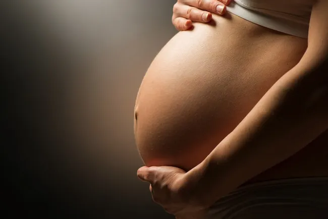
Pregnancy is often hailed as beautiful journey with lots of hope and wonder. Yet the human body can make something extraordinary and sometimes inexplicable, transformation. One such rare occurrence is lithopedion or stone baby. This phenomenon happens when decreased extrauterine foetus becomes calcified, forming a literal “stone child”. In intrauterine pregnancy the fertilised ovum implants and develops inside the uterus, which is ideal location for foetal development. Whereas, in extrauterine pregnancy the fertilised egg is implants and develops outside uterus.
The term lithopedion is
derived from a Greek works “lithos” means stone and “paidion” means child. It
refers a rare condition where dead foetus that developed outside the uterus
undergo calcification. Calcification is process where calcium ions build upon
the body tissue, causing it to harden. This process usually occurs in the
abdominal cavity following an abdominal pregnancy, an extrauterine
pregnancy. Normally, the process of
calcification in human body is very rare condition except in bone formation. This
process occurs when any portion of non-infected dead tissue too large to be
absorbed by the body will eventually undergo calcification.
For a foetus to form lithopedion, several conditions
must be met. First, the foetus must die due to unsuitable environment. Then,
the dead foetus should remain sterile to avoid decomposition and infection. It
needs to be retained in the abdominal cavity for at least 3 months to allow
sufficient time for calcification. Finally, condition favourable for calcium
ion deposit must be present. Lithopedions are classified into three types
according to kuchenmeister’s classification. First, lithokelyphos (stone sheath
or egg shell) where only the membrane surrounding the foetus are calcified. Second,
lithokelyphopedion (stone sheath child) in which both the membranes and the foetus
itself calcified. Finally, true lithopedion (stone baby) where the foetus is infiltrated
by calcium salt, with negligible membrane calcification.

Lithopedions are generally diagnosed incidentally during surgeries, autopsies, or through radiographic imaging of the abdomen or pelvis. Abdominal ectopic pregnancies are uncommon, and lithopedion are very rare. The calcified foetus can remain in the abdominal cavity for years without causing symptoms. However, there are instances where the foetal body parts are extruded through body orifices. Surgical removal is recommended once diagnosed to prevent further complication.
On analysing the statistical data on lithopedion depicts,
20% of foetus lived from three to six months, 28% between seven to eight months
and 43% nine months and term. The maternal age diagnosis was 23 to 35 in 27% in
some cases and 45 to 100 years in 63% of cases. In most of the case the
lithopedion was retained for four to twenty years. In some cases, it was
retained for 25 to 50 years.
While lithopedian is a rare and often devasting condition,
it’s important to remember that advancement in medical science continue to
improve our understanding of pregnancy complications. It essential to seek
medical attention without making delay, by raising awareness, we can work together
for earlier detection and prevent such tragic outcomes.
REFERENCES
- Moshiri M, Salari AA, Mansorian HR, Shariat R. Lithopedion (stone baby). Annals of Saudi medicine. 1996 Jan;16(1):69-70.
- D'Aunoy R, King EL. Lithopedion formation in extrauterine fetal masses. American Journal of Obstetrics and Gynecology. 1922 Apr 1;3(4):377-84.
IMAGE CREDITS


Comments
Post a Comment